Research
Plant cell biology with focus on integrated plant biology
We currently work on a number of different studies. Some examples are given below:
Sieve Tube Proteomics
Mature sieve elements contain four major components which are: mitochondria, P-proteins, sieve element plastids and ER. While the function of organelles in other major cell types is well understood, the function of the sieve element components is largely unknown. As mature sieve elements lack a nucleus, nucleic acid based omics approaches are of little help. We recently developed a protocol for the isolation of sieve tubes. Through proteomics we identified 124 proteins that are unique to sieve elements. In an NSF funded project we currently work on elucidating the function of those proteins. A publication in PNAS that describes the process and results was just accepted.
Phloem transport and the pressure flow hypothesis
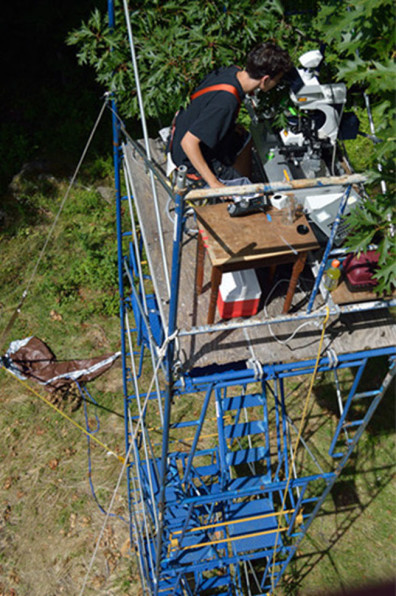
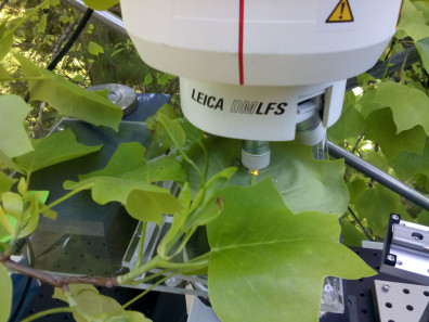
Energy rich molecules produced by photosynthesis feed our planet. The majority of these substances, however, are not consumed at the site of assimilation but are moved to distant tissues for growth, energy conversion, and/or storage. Photoassimilate translocation in plants takes place in the phloem and an estimated 90% of the food we consume has been translocated through this tissue. Therefore the phloem tissue is of fundamental importance for food security, impact of climate change on plant performance, and the development of bioenergy crops. Yet our understanding of the mechanism of phloem transport relies on a 85 year old , largely untested hypothesis. The complexity of the phloem tissue and the difficulties to access it for cell biological investigations has left us with major problems to understand even its basic functions such as the mode of translocation and allocation of assimilates. We currently study phloem cell biology and transport which requires taking microscopes into the canopy of trees for in situ studies.
Forisome Research
Some years ago we discovered the reaction of specific proteins in the phloem of bean plants which we called forisomes (gate bodies).
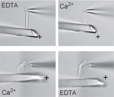
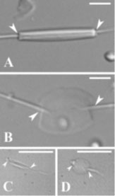
Forisomes serve as valves in the phloem to prevent leakage of photoassimilates after abiotic (mechanical) or biotic (e.g. aphids) injury. We have isolated forisomes and found that they have remarkable properties. Calcium ion concentrations above a threshold of 50 µM induce the reversible anisotropic deformation: a longitudinal shortening with simultaneous radial expansion which results in a drastic volume increase of up to 800% [see Figure 2 above]. In contrast to other motoric active proteins such as kinesin and myosin actuators, forisomes are completely independent of ATP.
In vitro, transformation of forisomes to a high volume state can also be triggered by pH changes in the range between pH 9.3 and 10.6. Since pH changes can be induced by amperometric ion titration, forisome reaction can be controlled electrically. Induction of more than 4200 contraction and expansion cycles did not indicate any decrease in reactivity. Moreover, forisomes generate significant forces during longitudinal contraction and expansion, which represents another unusual and useful property, since no antagonist is required (Figure left above).
The combination of useful properties renders forisomes paradigmatic models as actuators for (bio)-nanotechnological applications.
Root growth in dependence on soil water potential
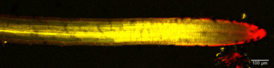
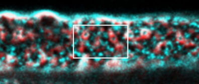
With changing climate, many soils on our planet become increasingly dry for longer periods. Plants follow several strategies to counteract unbeneficial conditions e.g. by higher water use efficiency or the induction of root growth into deeper soil layers. Yet, root growth requires water. We currently investigate how roots can grow in soils of varying water potential to understand the mechanisms behind growth control and adaption to changing environmental conditions. The first picture shows a single frame of a movie of a growing root tip in the soil. The movie was taken by Tim Ross-Elliott with a confocal laser scanning microscope (image left). Cell membranes are stained red and incoming water and solutes appear yellow.