Hair Cell Death, Protection, and Regeneration
How do hair cells die, and how can we prevent this damage and preserve hearing? This program examines the cell signaling pathways that are activated following exposure to different hair cells toxins such as aminoglycoside antibiotics, chemotherapy agents, and noise. We use the lateral line of larval zebrafish as a model system to study hair cell death and protection. Unlike humans, fish can regenerate their hearing, and we also use this system to understand the genetics of hearing regeneration. This research is funded by the American Hearing Research Foundation, American Otological Society, and Hearing Health Foundation.

The lateral line is an array of clusters of hair cells distributed along the head and body of the fish. Most fish have a lateral line system and use it to detect water movement near the fish-movement that signals a prey item nearby, the approach of a predator, or perhaps a mating opportunity. Hair cells in the lateral line are homologous (evolutionarily related) to the hair cells in our inner ears, and in fact hair cells in all vertebrate ears. In the picture above, each yellow dot is a neuromast (cluster of hair cells) labeled with the dye DASPEI.
We use a combination of drug screening and genetics to identify specific molecular players necessary for hair cell death in response to drug or noise damage. By understanding the intracellular death cascades that are activated by a specific toxic stimulus, we can then design therapeutic strategies to prevent this death process and preserve hearing.


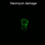
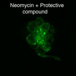
This pair of images show an individual neuromast from a transgenic fish where the hair cells are labeled with green fluorescent protein (GFP). The neuromast on the left is damaged after treatment with neomycin, while the neuromast on the right was co-treated with neomycin and a drug that prevents hair cell damage.
