Knowing SDS-PAGE gels
For in-gel digestion of proteins, basically any type of SDS-PAGE gel will work (gradient or non-gradient, self-cast or precast, reducing or non-reducing), as long as you use high-grade reagents to prepare them, and fresh buffers to run them. If you use home-made gels, don’t cast and run them on the same day. Leaving a gel overnight will allow polymerization to complete, thus preventing crosslinking of proteins in the gel by residual radicals. This is best done at room temperature (not in the fridge/cold room) by covering the gel in plastic, adding some wet tissue to prevent drying of the gel. Gels can be kept at least a week before running. Please observe the shelf-life of precast gels.
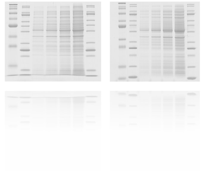
Coomassie-stained gels
Not all coomassie stains are compatible with mass spectrometry. We strongly recommend that you mix your own coomassie stains (recommended recipe). Always try to only stain your gel for the minimum time to (just) detect your protein(s) of interest. Remember that staining is only meant to visualize the band. Extended staining will increase the background in subsequent MS analysis, it will NOT increase the amount of protein. Destain the gel thoroughly to clear the background and to enhance visibility of the band. NEVER heat your gel in the microwave to speed up the staining! This will bake your protein in the gel, it will never get out again.
We DO NOT accept samples that have been stained with any commercial ‘instant’ or ‘ready-to-use’ staining kits. Please NEVER use overhead projector foils for gel scanning as this prevents the gel from being used for MS experiments.
Scanning of gel bands
It is very helpful to have a scan of the gel before processing it for MS analysis. To prevent contamination (see below), first seal the gel in plastic (foil and sealer and scanner are available in the Facility, which you are welcome to use). Only use a dedicated lab-scanner that is not used for other purposes (e.g. in the office). A gel scanner is available in the Facility – feel free to use them. Do NOT use overhead transparencies for scanning – this WILL introduce contaminating polymers that inhibit protease activity.
Cutting of gel bands
We prefer to receive intact gels and do the cutting ourselves (if possible, include a scan appropriately indicating your bands of interest). Alternatively, you can cut bands yourself, but please observe the guidelines (also read the section below on ‘contamination’). After cutting, gel bands can be kept for months at -20°C before MS analysis without adverse effect. For uniformity in sample preparation, it may be a good idea to prepare other bands from the same gel at the same time, even if you are not planning to send them now.
Contamination
Contaminants may be introduced at several steps during sample preparation. Some will go unnoticed, others will totally obscure all proteins. Therefore, minimizing contamination is essential.
Keratins
Keratins are omni-present and notorious contaminating proteins originating from skin, hair, dust, clothes, chemicals, etc. Total elimination is virtually impossible (and not necessary), but over-abundance will result in the repeated identification of keratins, and not your protein of interest. Most contaminating keratins are believed to originate from dust collected on the gel after running it. Therefore, a clean lab environment will certainly benefit the outcome of the MS experiment. If you don’t trust your lab is sufficiently dust-free, you are most welcome to run/cut your gel in our Facility. Alternatively, there are a couple of measures you can take. For starters, wear powder-free nitrile gloves at all times. Latex gloves often contain starch, and thus a lot of protein, so do not use these. Never touch the gel (or the liquid it is floating in) with bare hands. Use clean trays/containers that are only used for the purpose of gel staining (and not for e.g. blocking solutions for Western blots), and leave them closed with a lid during staining/storage. Don’t lean over gels, scratching your head how to interpret the band pattern. If you take the gel out of the container (e.g. for scanning, cutting), thoroughly clean the area with water (no soap!) or 70% ethanol. Always use fresh solutions to prepare and run gels, including sample loading buffer. Old solvents tend to collect dust – literally.
Other contaminating proteins
Other frequently observed proteins are BSA, immunoglobulins, and other serum proteins. These usually originate from cell culture media, and can be reduced by extensive washing of cell pellets.
Polymers
In a way, polymers are more serious contaminants than keratins, since they tend to stick to HPLC columns either ruining them, or at least causing a severe memory effect in subsequent LC-MS runs. On top of this, most polymers ionize more easily than peptides – so that even minute amounts can be deleterious for the experiment. The most frequently observed polymers are various forms of PEG, which are present in some plastics but also in soaps, hand-creams (wear gloves!) and detergents. It is not always easy to trace back the source of a polymer contamination, but we have had issues with some brands of pipet tips (TopLab), and pipet tips that have been autoclaved in-house. Do not use coated (‘low-binding’) eppendorf tubes, and avoid using soft plastics (Parafilm!). Furthermore, NO detergents should be present in samples to be submitted for MS, especially NP-40 and Triton X-100 have a bad reputation.
Source: Proteomics Core Facility – Sample Preparation – User Guide – EMBL